How the gut runs a local reflex
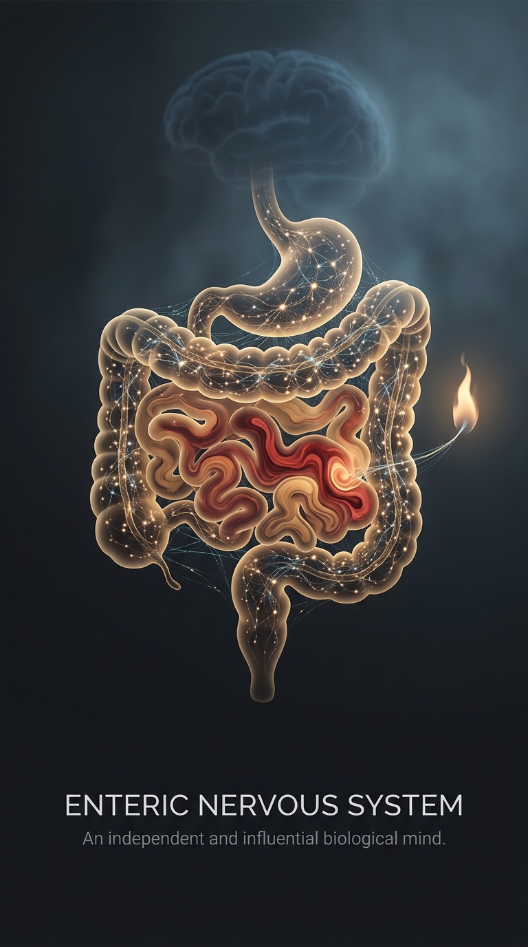
A meal sets off a chain of events the intestines can manage close to where they happen. Stretch sensors in the gut wall detect food and pressure; chemical sensors detect what is in the digestive tract. Enteric nerve circuits process those signals and activate nearby muscles and glands. The result can be a coordinated wave that moves contents along, timed with secretions that help break them down.
This local circuitry is arranged in networks within the gut wall. Some nerve cells sense conditions, others relay signals, and motor neurons act on muscle or glands. Because the connections form working reflex loops, digestion does not have to wait for a command from the brain for each contraction. The brain and spinal cord can still adjust the system—for example, through autonomic nerves that influence gut activity.
Serotonin works locally, too
Much of the body’s serotonin is produced in the gut, especially by specialized cells in its lining. There, serotonin helps digestive circuits respond to changes such as stretching or chemical irritation. It is a local signaling molecule, not a supply of serotonin sent straight to the brain: serotonin made in the gut generally cannot cross the blood–brain barrier, the protective boundary that limits what passes from blood into brain tissue.
The gut and brain nevertheless exchange information. Nerve signals can travel from the digestive tract toward the brain, while hormones and other chemical signals can also carry information between them. The connection is a two-way network, not a direct serotonin pipeline. That distinction helps explain how gut activity can influence sensations such as nausea or fullness without gut-made serotonin simply entering the brain.