How Cajal made individual neurons visible
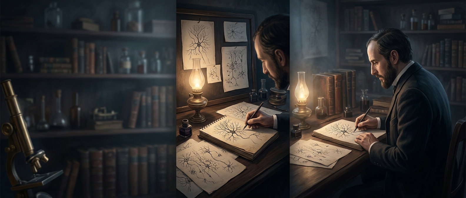
In the late 19th century, Spanish anatomist Santiago Ramón y Cajal used a staining method that colored only a small fraction of cells in a slice of brain tissue. Most cells stayed nearly invisible, while a few stood out in dark detail. That sparse contrast turned a tangled mass into something an observer could trace: a cell body with branching extensions reaching outward.
Cajal drew what he saw under the microscope, following those branches through crowded tissue. His illustrations made a crucial distinction clear: neurons—the nerve cells that carry information—were separate units, not parts of one continuous web. Their branches could approach one another closely without joining.
Why the gaps changed the picture
The tiny spaces between neurons are called synapses. A signal traveling along one neuron reaches its end, where it triggers a handoff to the next cell. In many synapses, that handoff uses chemical messengers that cross the gap and affect the receiving neuron. The cells therefore communicate across a boundary rather than passing a signal through one uninterrupted strand.
That separation gives the nervous system room to regulate communication. A receiving neuron can combine input from many connections, while the strength of particular connections can change. The same basic arrangement underlies circuits involved in sensation, movement, and memory.
Cajal’s drawings did more than make brain tissue easier to picture. By showing where one cell ended and another began, they helped establish the neuron as the nervous system’s basic working unit—and made the space between cells part of the explanation of how the brain works.