Hormones can alter the brain’s wiring
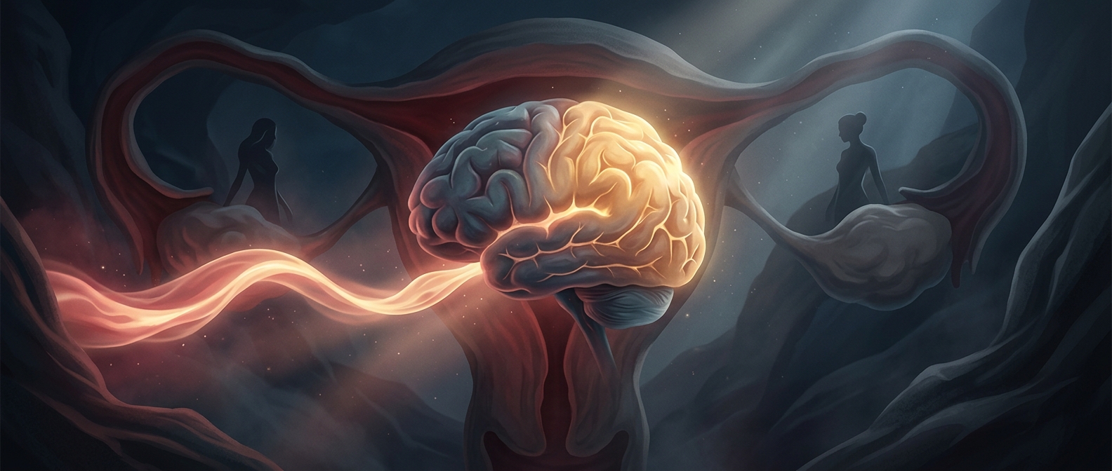
The brain is not a fixed structure that simply receives instructions from the rest of the body. Estrogen and progesterone can interact with receptors in brain tissue, influencing how neurons communicate and how connections between them are maintained. As hormone levels rise and fall during a menstrual cycle, these signals can shift the brain’s ongoing processes of adaptation, or plasticity.
That is one reason researchers have detected cycle-linked differences in the measured volume of some brain regions. A volume change on a scan does not mean that a region has gained or lost a chunk of brain. It can reflect subtle changes in tissue properties, including the connections and supporting structures that help neurons function.
A scan is not a mood or memory test
The hippocampus helps form and retrieve memories; the amygdala helps process emotionally important information. But finding a change in either region’s measured volume does not, by itself, show that someone’s memory or emotional control has improved or worsened. Brain structure, activity, and everyday experience are related, but they are not interchangeable.
The pattern can also differ from person to person and from one cycle to another. The useful takeaway is not that the brain becomes a different brain each month, but that reproductive hormones are part of the changing chemical environment in which it works. That makes the menstrual cycle relevant to brain research—not just to research on reproductive organs.