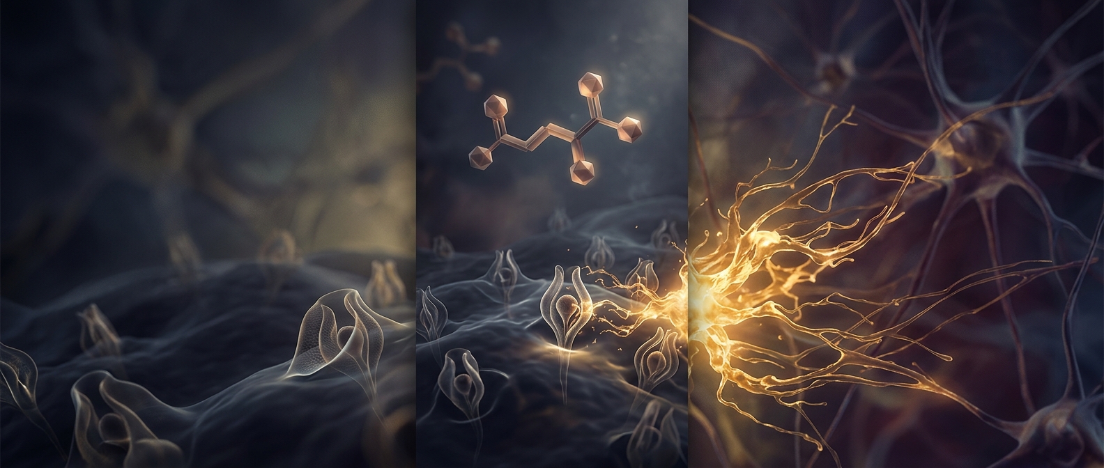
How Formaldehyde Locks Tissue in Place
A tissue sample is a crowded mesh of proteins, membranes, and other molecules. Once removed from the body, enzymes and microbes begin changing that mesh. Formaldehyde slows the damage by reacting with certain chemical groups on proteins. It can create bonds between nearby molecules, effectively fastening parts of the tissue’s microscopic framework together.
In laboratories, samples are commonly exposed to formaldehyde in a water-based solution called formalin. The solution must penetrate the tissue before it can preserve the interior, so a large piece may not fix evenly. Once the proteins are chemically connected, the tissue is less likely to soften or fall apart during handling and later preparation.
Preservation Comes With a Tradeoff
Fixation is not the same as freezing a specimen in its original living state. The process changes the molecules it is meant to preserve: proteins become less flexible, and some lose the shapes or chemical features that researchers might otherwise want to examine. That is why tissue preparation depends on the question being asked. A sample intended to show overall structure may be fixed, while tests of particular molecules may require different handling.
The practical payoff is that delicate arrangements—such as the layers of a small organ—can survive cutting into thin sections for a microscope. Formaldehyde preserves a physical map, not life itself: the cells are no longer functioning, but their connected structure is sturdy enough to inspect.