How a tiny current helps set the heart’s direction
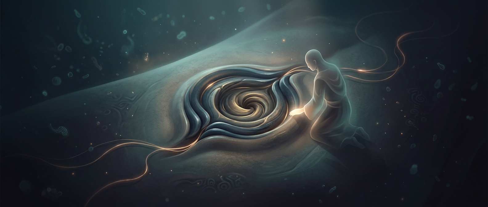
Early in development, an embryo has to distinguish left from right before organs take their places. Near its midline is a small region containing cilia—microscopic, hairlike structures. As they rotate, they create a gentle flow of fluid toward the embryo’s left. Cells detect that directional cue, helping establish different molecular signals on the two sides.
Those signals give developing organs a shared orientation plan. They do not physically push the heart into place; they influence how its cells grow and arrange themselves.
The heart turns as it forms
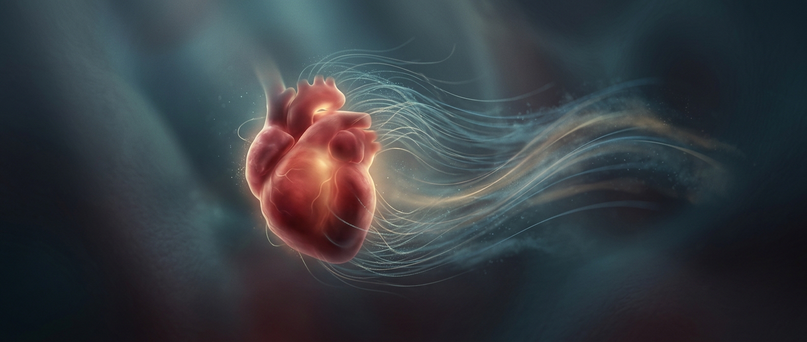
The heart first develops as a roughly straight tube. As it lengthens, it bends and loops in a consistent direction, bringing its future chambers and major vessels into their characteristic arrangement. Because this turn happens while the heart is taking shape, its position is built into its developing structure—not the result of a finished organ sliding across the chest.
The cilia-driven flow is an important early cue, but it is one part of a larger chain of development. If left-right patterning takes a different course, the heart and sometimes other organs can form in a mirror-image arrangement. That possibility reveals the key point: the embryo must set a direction early, before a heart exists that could simply be moved.