How brain scans can hint at a dream’s images
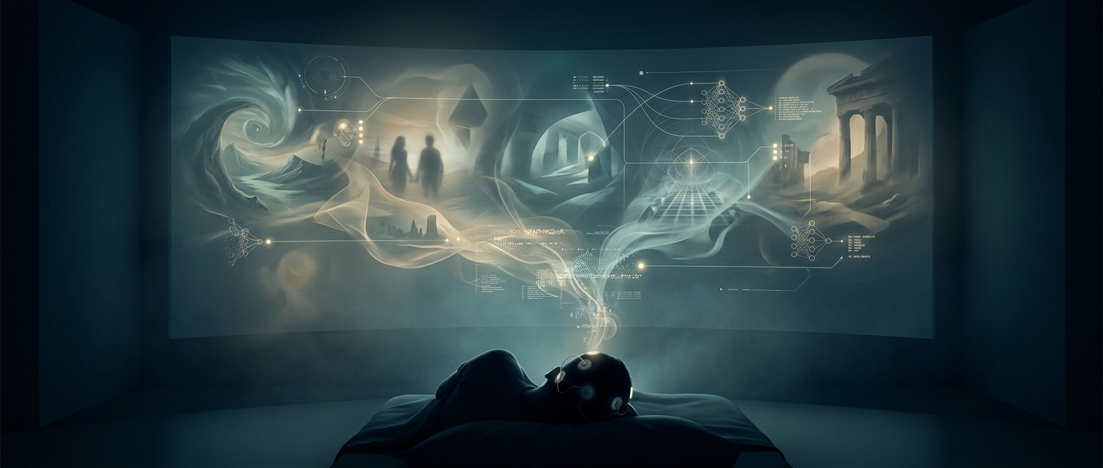
An fMRI scanner does not photograph thoughts. It tracks changes in blood oxygen that accompany brain activity, including activity in visual regions while a person sleeps. Those signals are too coarse to reveal a crisp picture on their own, but they can contain patterns associated with broad visual features—such as shapes, textures, or categories of objects.
To make those patterns useful, researchers first compare a person’s brain responses with images the person sees while awake. A computer learns which signal patterns tend to accompany which kinds of visual content. During sleep, it can then use the learned associations to generate an image that fits the recorded activity.
Why the result is a reconstruction, not a replay
The system is making an informed guess from noisy, indirect evidence. It does not capture a complete dream or recover exactly what the sleeper saw in their mind. The resulting images can be blurry yet recognizable because the model can identify likely visual elements without knowing every detail of the imagined scene.
Dream reports help researchers check whether the output broadly matches what a person remembers after waking. But memory is imperfect, and a match to a reported object does not prove that the machine has reproduced the dream exactly. The images are best understood as visual interpretations of brain data, not recordings of private experience.
That distinction is what makes the technique striking: even an imperfect signal from a sleeping brain can carry enough structure for a computer to turn some of its visual content into something people can inspect.