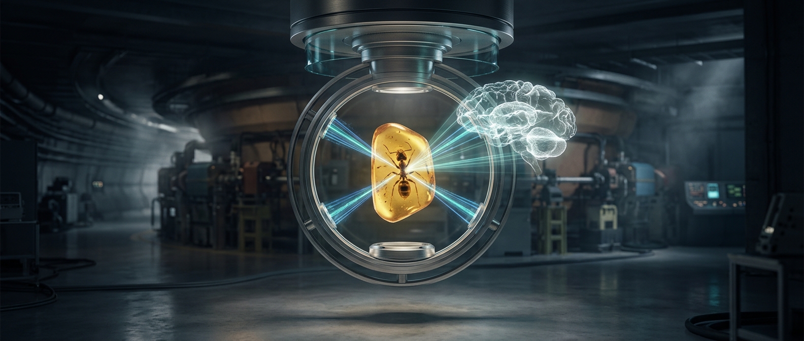
Reading an insect fossil without opening it
Amber can preserve an insect in remarkable detail, but the resin surrounding it also hides the animal’s interior. Cutting the piece open would risk destroying both the specimen and its scientific value. Instead, researchers use X-ray computed tomography, or CT: the same basic imaging approach used in medical scanners, adapted to fossil material.
From overlapping shadows to a 3-D brain
As X-rays pass through the amber from different angles, denser and less-dense materials absorb them differently. The insect’s body, surrounding resin, and tiny internal structures therefore leave slightly different patterns in the scans. A computer combines those measurements into a three-dimensional volume. Researchers can then move through it as a sequence of virtual slices, separating structures that overlap in an ordinary photograph.
That distinction matters for a brain, which is soft tissue and does not survive as living tissue in a fossil. What imaging can reveal is its preserved shape and the outlines or impressions left by its structures—not a functioning brain or its original cells. The resulting anatomy can still help scientists compare the insect with living relatives and investigate how its body was organized.
The scan has limits, but keeps the specimen whole
The view depends on image resolution and on how clearly the insect differs from the amber around it. Very small features may blur together, and preservation can obscure or distort anatomy. Even so, scanning makes it possible to study hidden structures without physically exposing them. The amber remains intact, while researchers gain a view that a surface photograph could never provide.