How the body’s left and right can switch
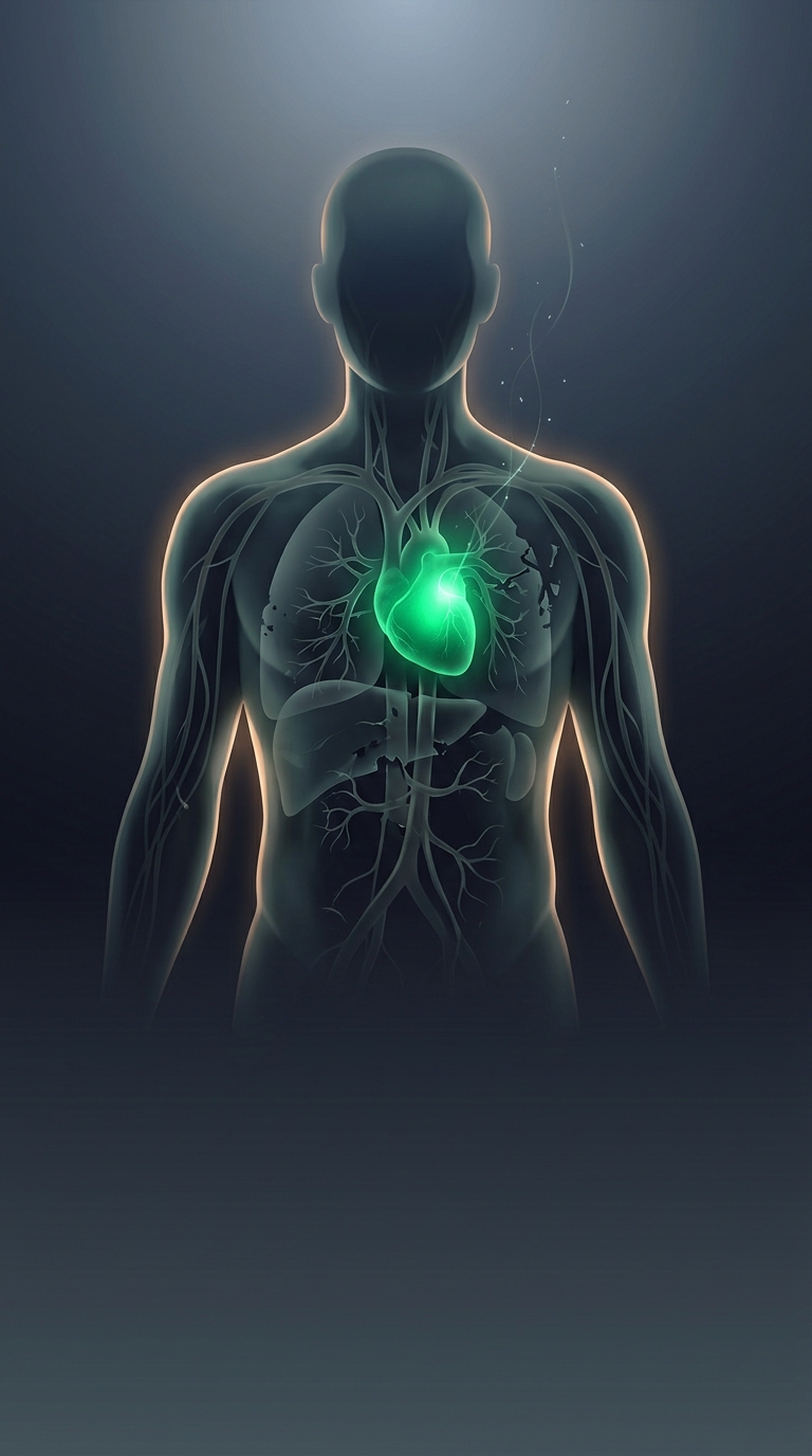
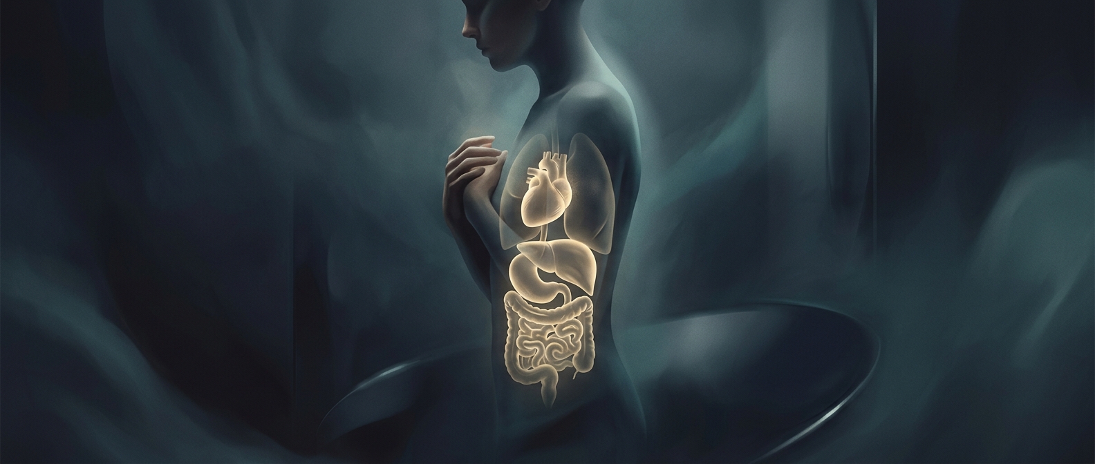
Early in development, an embryo establishes which side will become the body’s left and which will become its right. In situs inversus, that left-right pattern is reversed for major organs: the heart may point right, while the liver and stomach also occupy positions opposite their usual ones. The arrangement is often called a mirror image because the organs’ positions are reversed across the body’s midline.
The switch happens as the body is being organized, not because the organs later migrate across the chest or abdomen. The exact developmental cause can vary; in some people, problems with tiny hairlike structures called cilia—which help generate signals involved in setting left and right—play a role.
Why a reversed layout may work normally
Organs can often function well in their reversed positions. Their tissues and connections develop as a coordinated arrangement, so a heart on the right is not automatically a failing heart. Situs inversus is also different from a defect in how an organ works, though some people have associated health conditions.
The unusual layout matters most when someone needs medical care. A clinician feeling for the liver, interpreting an image, or placing an instrument cannot assume the usual landmarks. Recognizing the reversed anatomy helps prevent confusion and makes procedures safer. In some cases, it is found incidentally when imaging for an unrelated reason reveals that the body’s internal map runs in reverse.