Mapping the brain without putting the patient to sleep
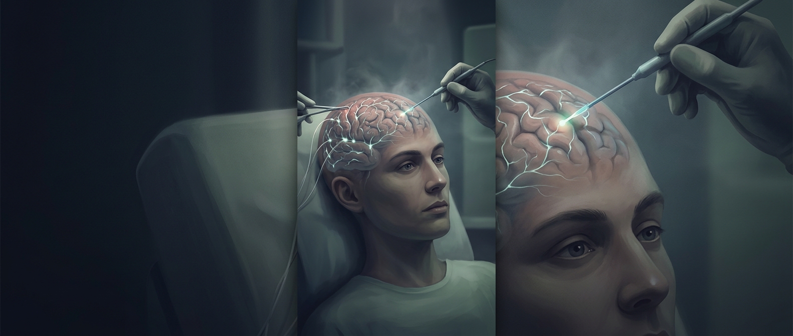
Penfield used electrical stimulation during operations for epilepsy, when surgeons needed to identify abnormal tissue without damaging the brain areas patients relied on for movement and sensation. Keeping a patient awake for parts of the procedure made it possible to check those functions in real time. The brain itself does not feel pain, though the operation required careful preparation and the patient’s cooperation.
A surgeon could touch an exposed area of the brain with a small electrode and apply a brief current. If the patient reported a sensation or showed a movement, the response offered evidence about what that patch of tissue did. Repeating this across nearby points turned the operation into a practical test: stimulate, observe, and mark the result.
A body map with uneven proportions
The resulting map was not a neat outline of the body. Areas involved in precise or highly sensitive functions—such as those of the hands and face—occupied more brain territory than their physical size would suggest. This distorted body map, often called the cortical homunculus, reflects how much processing different body parts demand, not how large they are.
The map also helped surgeons make decisions under a difficult constraint: removing tissue could reduce seizures, but taking too much—or cutting through a critical functional area—could leave lasting problems. Stimulation did not reveal a complete, fixed blueprint of the mind; it provided a direct, patient-specific guide to important functions near the surgical site.
That practical use is what made the map more than a scientific curiosity: a brief twitch or reported tingle could help determine where a surgeon should stop.