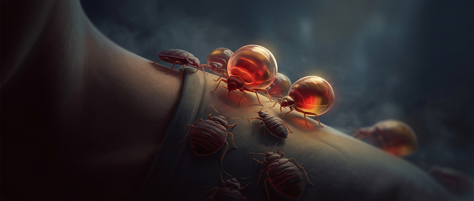
How a Tick Builds a Grip in Your Skin
A tick’s feeding tube is not a smooth needle. Its central mouthpart, called the hypostome, is a narrow, barbed structure: backward-facing points help resist being pulled out. Around it, the tick secretes saliva that sets into a firm sheath, anchoring the mouthparts in the small wound.
This “cement” is made from proteins and other substances in the tick’s saliva. It is not household glue; it is a biological material that hardens at the bite site. The barbs and the sheath work together, spreading the job of holding on across both the shape of the mouthpart and the material around it.
The Bite Becomes a Feeding Site
Once attached, the tick feeds slowly, drawing blood and tissue fluids through its mouthparts. Its saliva helps keep the feeding site usable: it can interfere with clotting and dampen local immune and inflammatory responses. That matters because a host’s normal defenses would otherwise make the wound less hospitable, swelling it or sealing it with a clot.
The attachment is therefore more than a quick puncture. The tick establishes a small, stable feeding site and maintains it while feeding, which can last for an extended period depending on the tick and its stage of development. The hardened sheath helps keep the mouthparts seated as the tick remains in place.
Why the Grip Matters
A barbed mouthpart alone could still shift or tear at the wound as the tick feeds. The cement-like material reinforces the connection, making prolonged attachment more reliable. It is a striking example of how a parasite can use both anatomy and secretions: the tick supplies the structure, then builds support around it—right at the point where it needs to feed.