How the eye turns light into a retinal image
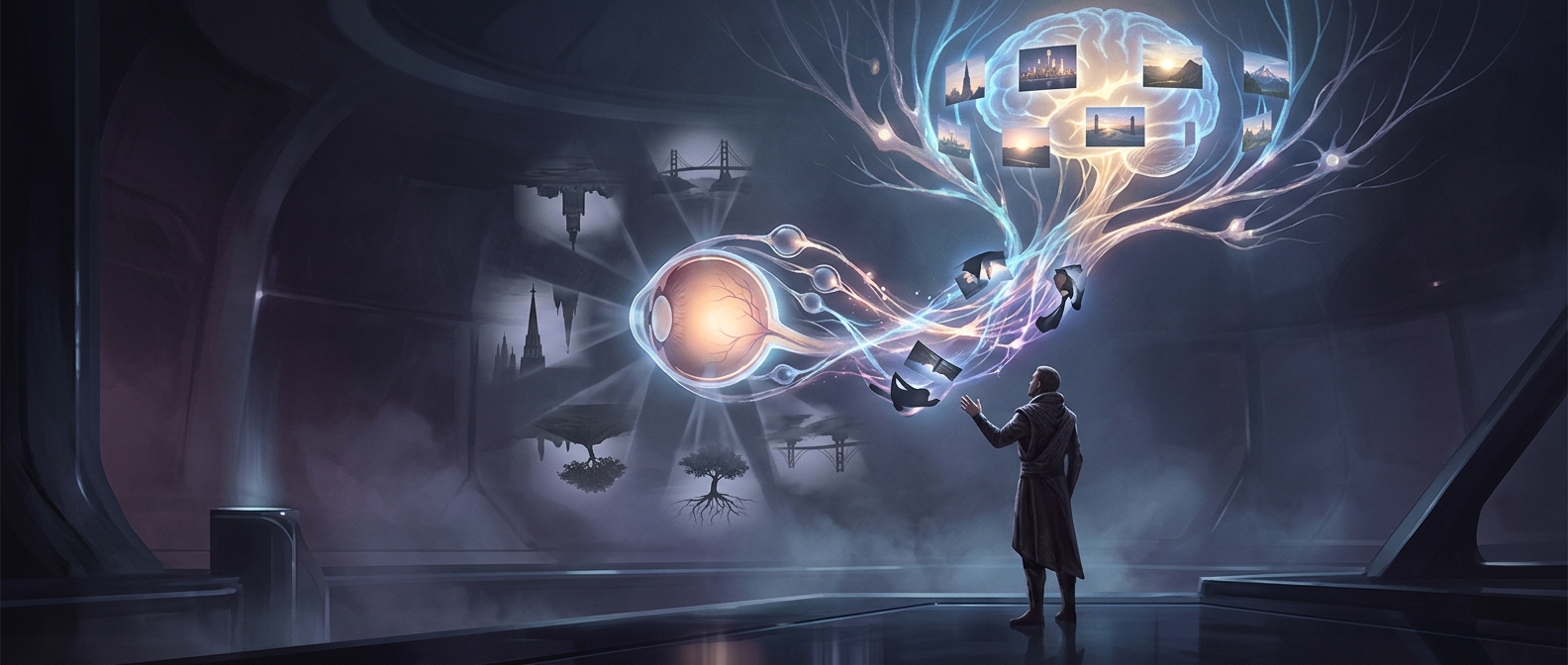
The cornea—the clear front surface of the eye—and the lens bend incoming light so it converges on the retina, the light-sensitive tissue lining the back of the eye. Rays from the top of a scene cross on their way through the eye and land lower on the retina; rays from the bottom land higher. The result is an inverted optical image, much like the image formed by a simple camera lens.
The retina is not a screen that sends a tiny picture to the brain. Its cells respond to light and convert it into electrical signals. Those signals travel along the optic nerve to several brain regions that process visual information, including areas that help identify edges, motion, color, and position.
Why the world does not look upside down
The brain does not need to rotate a mental photograph. Instead, it interprets patterns of activity from the retina in relation to the visual system’s organized connections and to other information, such as eye movements and body position. Through this processing, the lower part of the retinal image corresponds to what we perceive above, and the upper part to what we perceive below.
That distinction matters: the image is inverted in the eye, but perception is not a picture the brain flips over. Vision is the brain’s ongoing interpretation of signals, not a view of the retina itself. The correction is therefore not a conscious step—it is built into how the visual system makes the world’s positions meaningful.