Why the Motor-Cortex Map Enlarges the Hands
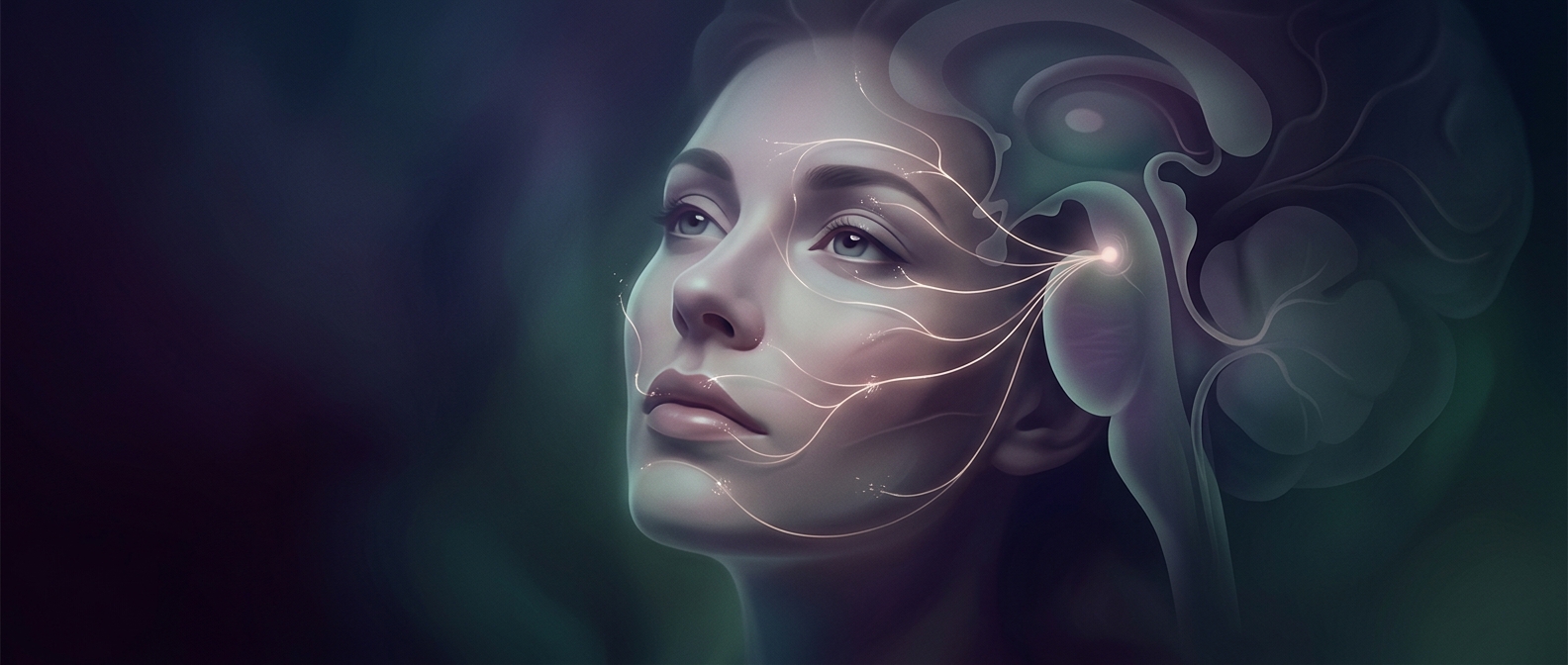
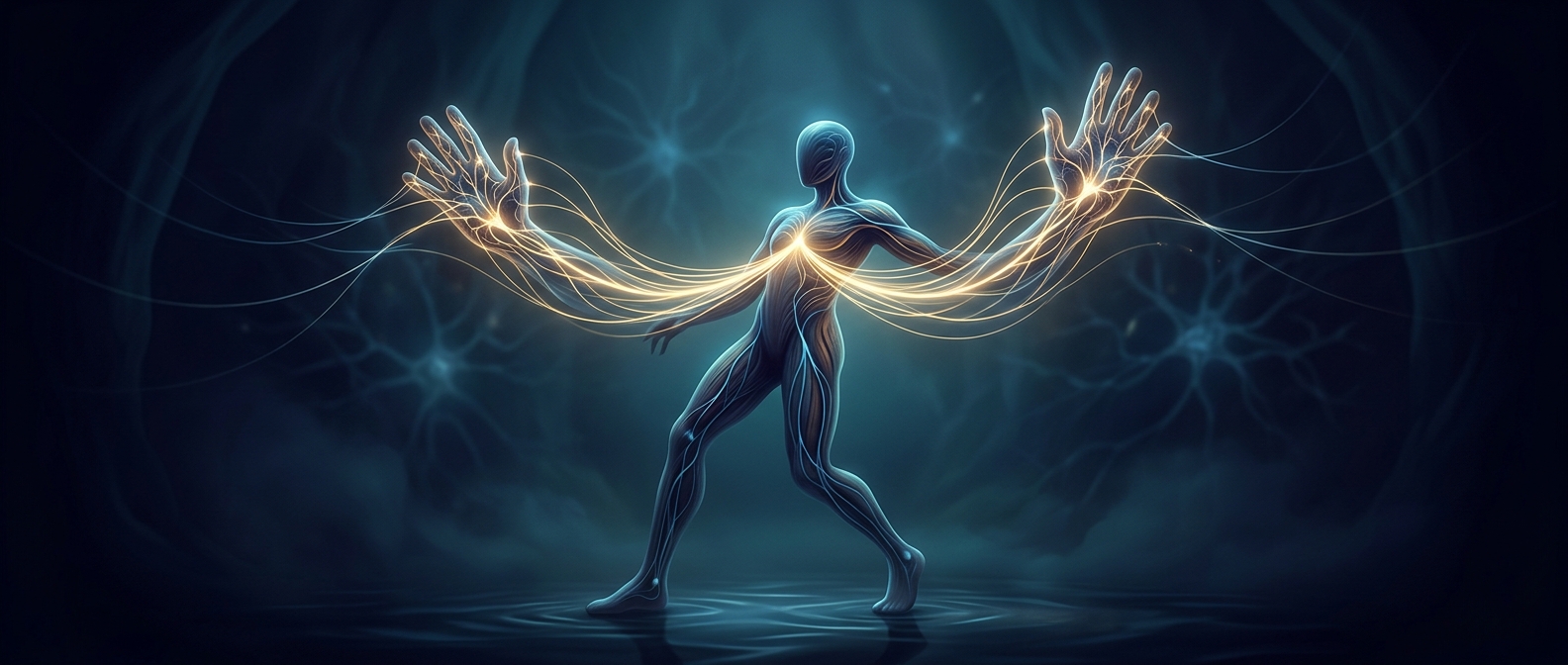
The brain’s movement map is not scaled to body size. In the primary motor cortex—a strip of tissue near the top of the brain that helps send voluntary movement commands—the regions involved in controlling the hands and face occupy unusually large areas. This arrangement is often illustrated as a distorted figure called the motor homunculus.
That distortion reflects the complexity of the commands, not the physical size of the body part. A hand can reach, grip, pinch, and adjust pressure through many combinations of muscles. Picking up a paper clip, for example, requires the fingers to move in a coordinated pattern while making tiny corrections so the object does not slip. The cortex devotes substantial circuitry to producing and refining these varied movements.
A Map, Not a Set of Isolated Buttons
The drawing can make it seem as if each body part has a neat, separate patch of cortex. In reality, neighboring regions overlap, and movement depends on networks of brain areas working together. A cortical map is a useful guide to broad organization, not a literal diagram in which one small spot controls one finger and nothing else.
The map is also adaptable. With practice, the brain can change how movement-related circuits respond, helping a person perform a skilled action more efficiently. But the basic imbalance remains revealing: brain space is allocated less by how much of the body a part occupies than by the range and precision of control it demands. That is why a hand can take up far more room in a brain diagram than its modest size would suggest.