Why the Retina Has a Blind Spot
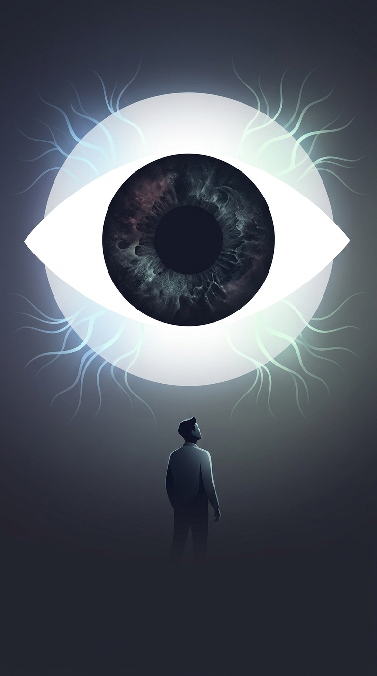
The eye’s blind spot is a small patch of the visual field where light cannot be detected. It occurs at the optic disc, the place where the optic nerve carries signals out of the eye and blood vessels enter. At that point, the retina—the light-sensitive layer lining the back of the eye—has no photoreceptor cells to register an image.
To find the spot, close one eye and fix the other on a small mark. Move a second mark slowly across a plain background. At a particular distance, the moving mark can disappear when its image falls on the optic disc. The effect is easier to notice with a uniform background because there are fewer surrounding details to reveal the gap.
Why Vision Still Looks Continuous
The brain does not receive a complete picture from either eye. Instead, it combines signals from both eyes and uses nearby visual information to fill in missing areas. With both eyes open, one eye can usually supply information about a scene that falls in the other eye’s blind spot. Even with one eye closed, the brain often extends a surrounding color or pattern across the gap.
This filling-in is not a conscious repair or a literal reconstruction of every hidden detail. It is one way the visual system makes a stable, useful perception from incomplete signals. That is why the blind spot is normally hard to notice, even though it is present in each eye.
An Evolutionary Tradeoff, Not a Broken Eye
In the human retina, nerve fibers lie in front of the light-sensitive cells and gather at the optic disc to form the optic nerve. That arrangement creates an exit point with no photoreceptors. In an octopus eye, the nerve connections run behind the light-sensing cells, so the optic nerve does not pass through the retina in the same way and there is no equivalent blind spot.
The contrast shows that eyes can solve the same basic task with different wiring. Human vision works remarkably well, but its seamless appearance partly depends on the brain smoothing over a physical gap in the retina.