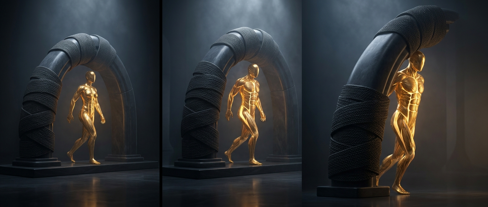
A sleeve woven from neighboring muscles
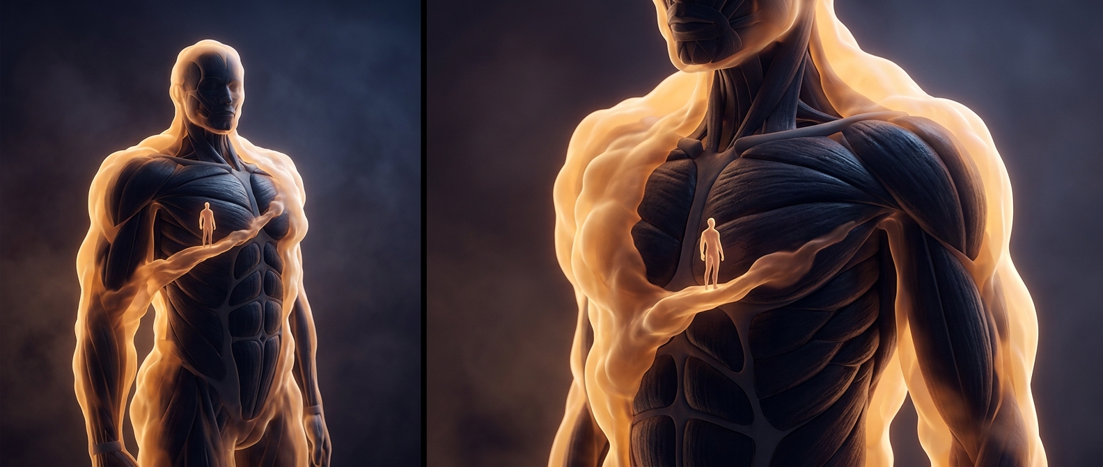
The rectus sheath is not a separate tube that forms on its own. It is built from broad sheets of connective tissue, called aponeuroses, that extend from the abdominal muscles on either side. These sheets pass in front of or behind the rectus muscles—the paired vertical muscles often called the “six-pack”—and meet at the midline in a tough seam called the linea alba.
This arrangement anchors the muscles while letting the abdominal wall flex and brace. When the rectus muscles contract, their force is transmitted through the sheath and its midline seam, helping the trunk bend and the abdomen resist pressure.
The sleeve changes lower down
For much of the abdomen, the rectus muscles have a connective-tissue layer both in front and behind them. Below a boundary called the arcuate line, the sheets from the side muscles pass in front of the rectus instead. Behind it, the muscle lies against a thinner layer of connective tissue and the lining of the abdominal cavity.
That change matters because the sheath is not equally thick in every spot. It also provides a route for blood vessels and nerves that supply the abdominal wall. Surgeons take these layers and pathways into account when planning incisions: the same muscle can be approached through different tissue arrangements depending on where the incision is made.