How a retinal pattern becomes an identity map
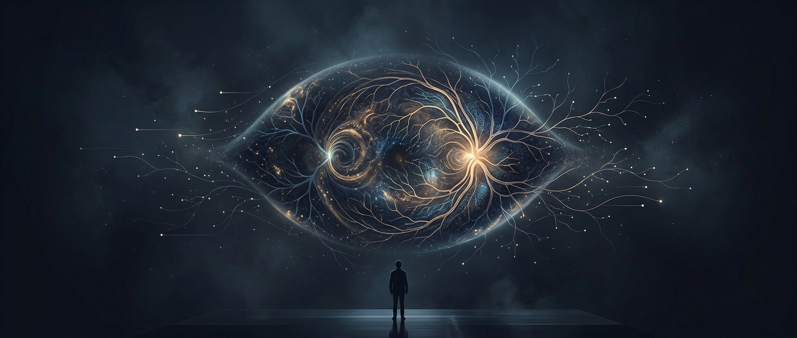
At the back of the eye, blood vessels divide and branch across the retina, the light-sensitive tissue that sends visual signals to the brain. A retinal scan records this network and turns its geometry into measurable landmarks: where vessels split, how they curve, and how branches relate to one another. The 256 identifying features are not 256 separate marks stamped onto the eye; they are details in a connected pattern.
From light to a usable pattern
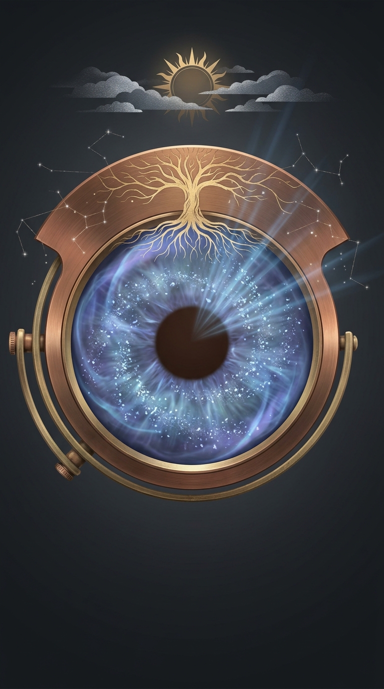
To capture the network, a scanner directs light into the eye and records the reflected image of the retina. The device must line up with the pupil and focus on the back of the eye, so the person usually looks steadily into the scanner for a moment. Software then analyzes the vessel layout and converts it into a compact representation that can be compared with a stored one.
That comparison is about the arrangement of the vessels, not simply their color or the overall appearance of the eye. A fingerprint reader looks for ridges at the fingertip; a retinal system looks for branching geometry deep inside the eye.
Why the pattern can identify someone
Blood-vessel networks form through growth, branching, and changing demands for blood supply. Their many turns and junctions create a detailed map that is difficult to reproduce in the same arrangement in another person. The result is a biometric identifier—a bodily pattern used to recognize or verify identity.
Because the map is inside the eye, capturing it requires a closer, more deliberate scan than photographing a face. The distinctive feature is also a reminder that an eye image can reveal more than what someone looks like: it can contain a structured biological pattern that serves as an identity key.