How a membrane turns a signal into a current
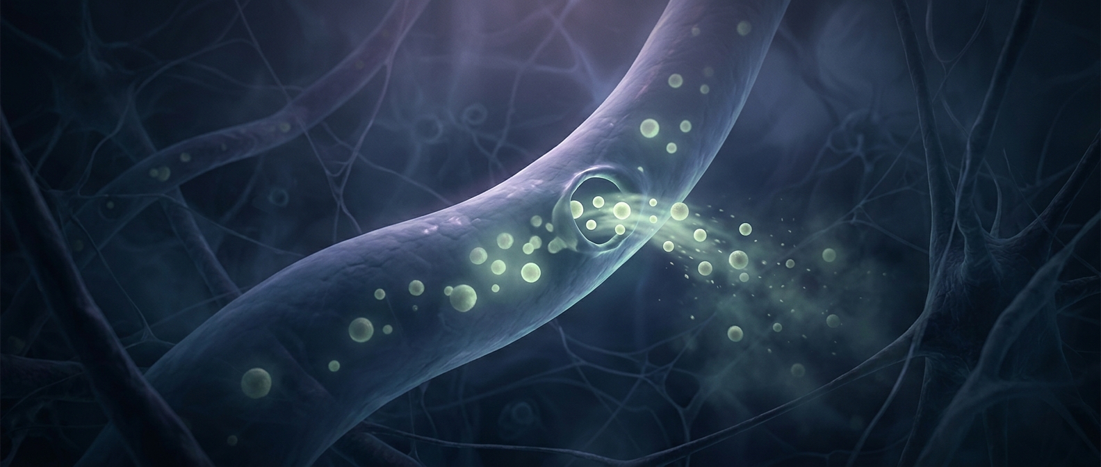
The fatty membrane that surrounds a cell is easy for some molecules to cross but blocks most charged particles. Ions—atoms or molecules with an electrical charge—cannot simply slip through its oily interior. Cells solve this with ion channels, proteins that form narrow, water-filled passages across the membrane.
A channel’s gate responds to a particular kind of change. Some open when the voltage across the membrane shifts; others respond when a chemical messenger binds to them, or when the membrane is stretched. The signal changes the protein’s shape, opening a route through the barrier.
Selective gates, driven by gradients
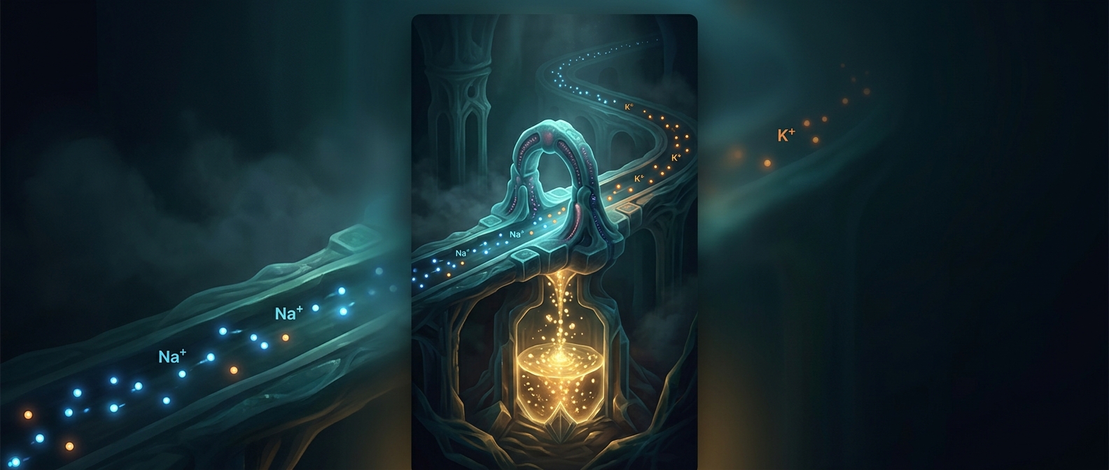
An open channel is not a pump. It does not spend energy to push ions through. Instead, ions move down an electrochemical gradient: the combined effect of differences in ion concentration and electrical charge on either side of the membrane. The channel’s shape and charged interior favor certain ions, so a potassium channel, for example, does not serve as an unrestricted passage for every charged particle.
That selectivity matters because even a brief opening can alter the cell’s electrical state. As ions cross, the balance of charge across the membrane changes. The channel then closes, limiting the flow; other channels and energy-consuming pumps help restore the original ion distributions.
A small opening, a large consequence
In nerve cells, coordinated channel openings help create the electrical signal that travels along a nerve. At a nerve ending, that signal can trigger the release of chemical messengers, passing information to another cell. In muscle, related electrical changes help set contraction in motion.
The “bouncer” image is useful, but the gate is not checking credentials consciously: its structure makes some ions more likely than others to pass, and its trigger determines when passage is possible. A tiny protein opening can therefore turn a local signal into a rapid change in what the whole cell does.