How three signals become millions of colors
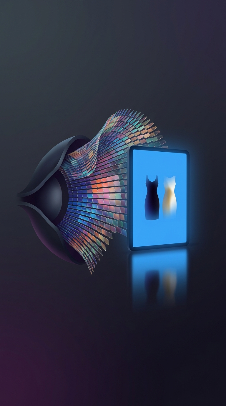
Color vision begins in the retina, the light-sensitive tissue at the back of the eye. Its three types of cone cells respond most strongly to different ranges of light wavelengths: broadly, long, medium, and short wavelengths. These ranges overlap, so a single patch of light can activate more than one cone type at once.
The brain does not receive a label such as “turquoise” from the eye. It compares the cones’ activity—how strongly each type responds relative to the others—and interprets that pattern as a color. A small shift in the balance can make a surface look bluer, greener, or more muted, even when the change in light is subtle.
Why the combinations multiply
Each cone type can respond at many levels, not simply switch on or off. The three streams of activity therefore form a large range of possible patterns. The brain further compares signals in ways that help distinguish differences in hue and brightness. Together, these steps let people with typical color vision tell apart millions of color variations under suitable viewing conditions.
That ability has limits. The same object can produce different cone signals under different lighting, and nearby shades may be hard to separate when they are dim or nearly identical. The brain uses surrounding colors and context to help interpret what it sees, which is why a color can appear to change when the light or background changes.
The striking point is that color is not a separate signal for every shade: millions of perceived variations emerge from the brain interpreting three overlapping channels.