A living view of the circulation
The retina—the light-sensitive layer at the back of the eye—needs a steady supply of blood to work. Its arteries and veins are unusually useful to clinicians because they can be viewed directly through the pupil, without an incision. With a bright light and a viewing instrument, an eye professional can inspect the vessels’ width, shape, and surrounding tissue.
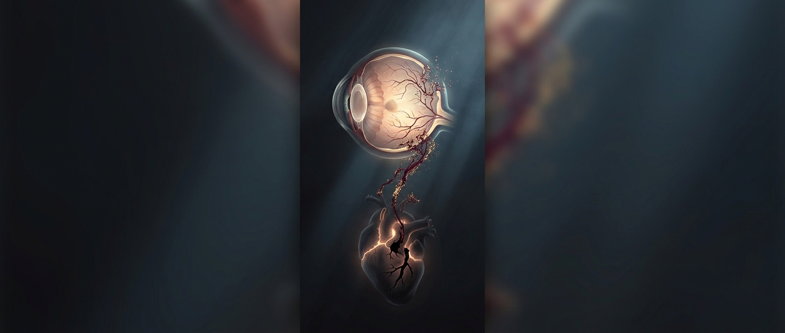
That view offers a small window onto a much larger network. The vessels in the eye are affected by forces that act throughout the body, including persistently high blood pressure and diabetes. Over time, these conditions can alter vessel walls or injure delicate retinal tissue. Signs such as narrowed vessels or tiny areas of bleeding may therefore point to problems that deserve attention beyond the eye.
A clue, not a verdict
Retinal findings do not reveal exactly what is happening in the heart, and one eye examination cannot determine a person’s future risk by itself. The same visible change can have more than one explanation, while cardiovascular disease may be present without obvious retinal signs. Clinicians interpret what they see alongside factors such as blood pressure, medical history, and other test results.
The practical value is in connecting two parts of health that can seem unrelated. A routine view of the back of the eye may raise a reason to check or better manage a risk elsewhere in the body. In that sense, the retina is not a substitute for a heart evaluation; it is a place where the circulation can sometimes leave an early, visible trace.