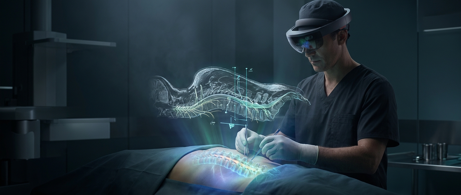
Turning a Scan Into a View Through the Body
A CT or MRI scan is a stack of thin images. By combining those slices, software can build a three-dimensional model of the patient’s anatomy, separating structures such as vertebrae and blood vessels so they can be viewed together rather than one slice at a time.
The HoloLens displays that model as an augmented-reality overlay: a digital image placed in the wearer’s view of the real world. When the model is aligned with the patient’s body, the surgeon can look at the back and see where hidden anatomy lies beneath the skin. Moving the head changes the viewpoint, much as it would when looking around a physical object.
Why Alignment Matters in Surgery
The useful part is not simply making a spine model appear in the room. The model must be registered—matched in position and orientation—to the patient on the operating table. The system described uses scan data to create a millimeter-accurate 3D representation, making the overlay a visual guide to structures the surgeon cannot see directly before an incision.
That view can help a surgeon understand the route to a target and the nearby anatomy before cutting. It does not replace the patient’s body with a digital one or perform the operation: the surgeon still interprets the image and makes the decisions. The practical promise is a more direct connection between what a scan shows on a screen and where that anatomy sits in the patient.