What brain scans can—and cannot—show about burnout
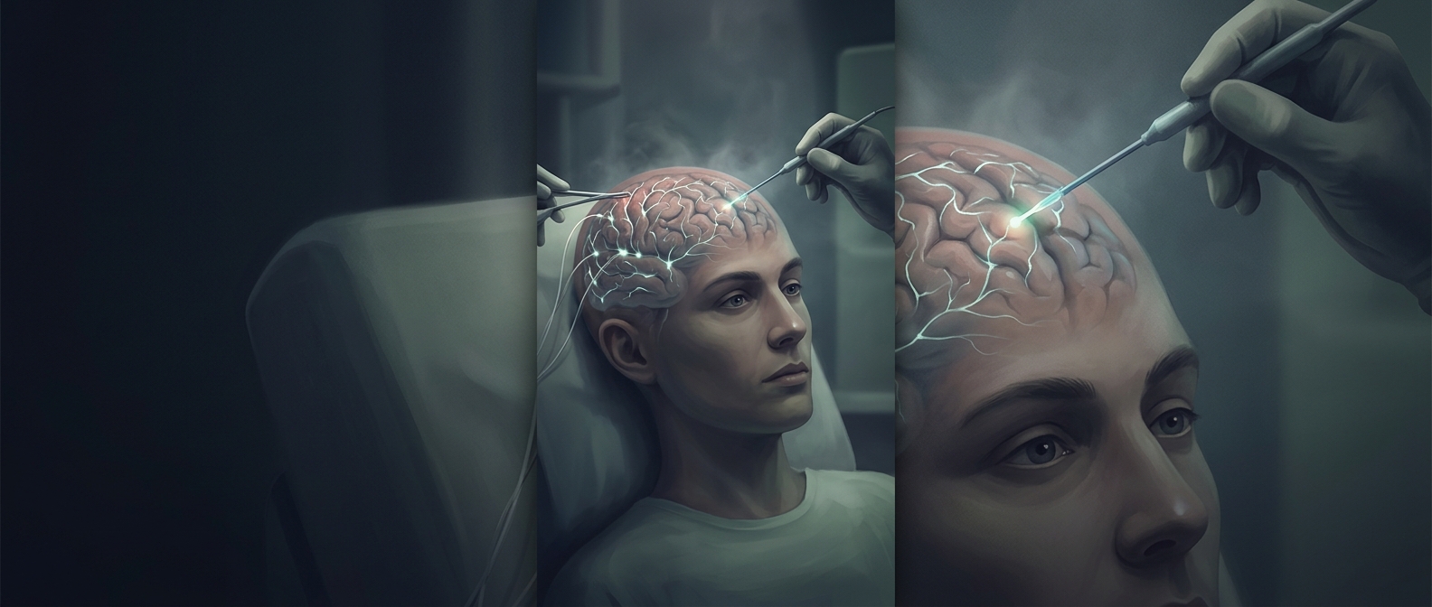
Burnout is more than feeling tired after a difficult shift. Under prolonged work stress, the brain’s systems for handling emotion, attention, and memory may adapt to the demands placed on them. Imaging research has reported differences in the hippocampus and amygdala among healthcare workers experiencing severe burnout. These are group-level findings, not a brain scan that can diagnose burnout in an individual.
How prolonged strain may leave a trace
The hippocampus helps bind experiences into memories and place them in context; the amygdala helps detect emotionally important or threatening cues. When stress is persistent, the body repeatedly activates systems that prepare it to respond to demands. That ongoing load may influence how brain regions function or appear on scans. But an observed difference does not, by itself, reveal the precise process that produced it.
There is also a timing problem: a scan captures the brain at one point, while burnout develops over time. If researchers find a difference in people with burnout, they cannot automatically tell whether work stress caused it, whether an earlier difference made stress harder to manage, or whether other factors contributed. Nor does an imaging difference necessarily mean lasting injury.
Why the distinction matters
The findings make burnout a concern that reaches beyond mood or motivation, but they do not mean every exhausted doctor has brain damage. They point instead to a practical possibility: sustained strain may affect the systems clinicians rely on to remember details and respond to emotionally charged situations. A brain image can offer clues about that burden; it cannot substitute for understanding a person’s symptoms, working conditions, and recovery.