How light made the retina visible
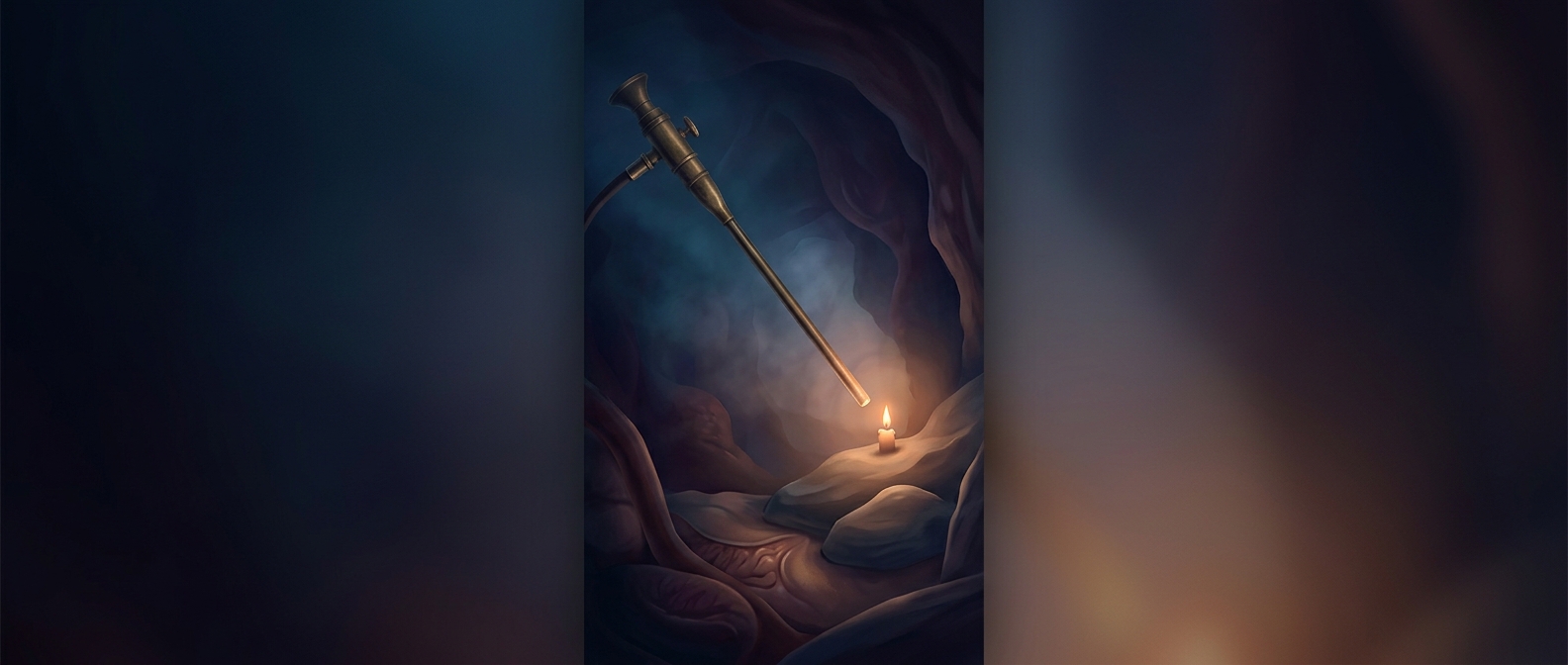
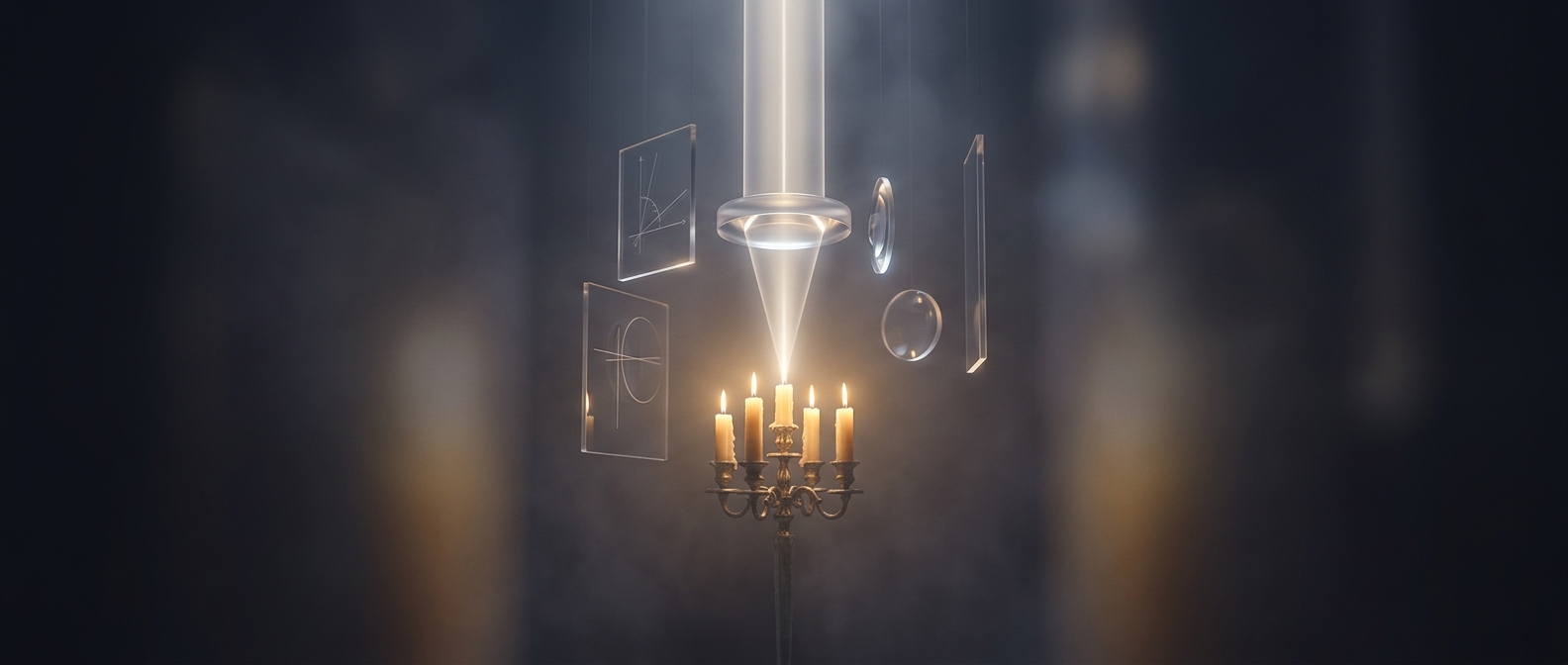
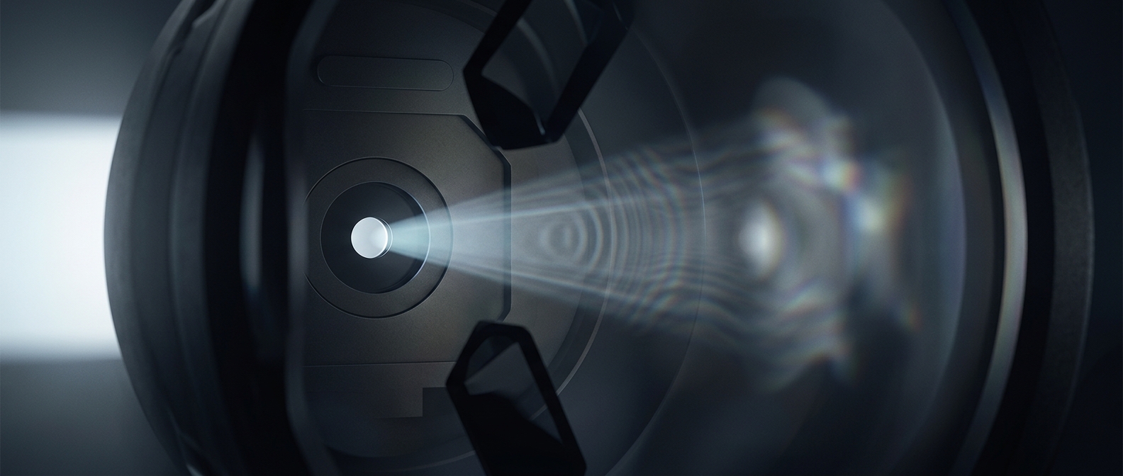
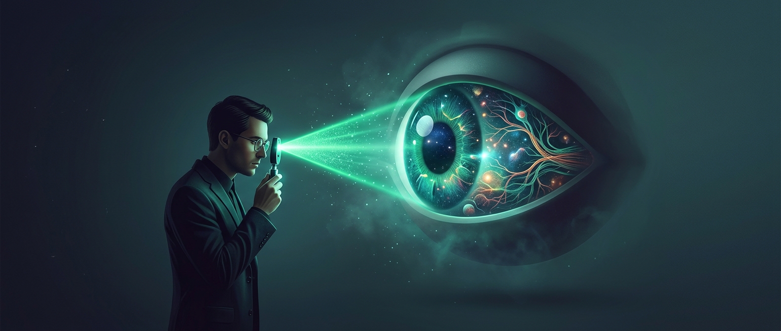
The retina sits at the back of the eye, but it is hard to see from outside: light must pass through the pupil and the eye’s clear focusing structures, then return along nearly the same route. Helmholtz’s instrument solved the alignment problem. It sent light into the eye while giving the observer a way to look back through the pupil toward the illuminated retina.
The crucial detail is that the pupil can serve as both doorway and viewing path. When the returning light reaches the observer’s eye, the retina appears as a lit surface rather than a hidden interior. The instrument does not open the eye; it uses the eye’s own transparent parts as a viewing channel.
A view with practical consequences
Once the retina could be examined in a living person, clinicians could inspect structures such as the optic nerve’s entry point and the branching blood vessels. Their appearance can offer clues about problems inside the eye, and sometimes about broader conditions affecting the body. The ophthalmoscope turned an internal surface that had been inaccessible without surgery into something a doctor could directly observe.
That change was more than a brighter view. It made examination repeatable: a clinician could direct the light, focus on a particular region, and compare what was visible across patients or over time. A small instrument thus made the back of the eye part of ordinary clinical observation.