Why cartilage repairs itself slowly
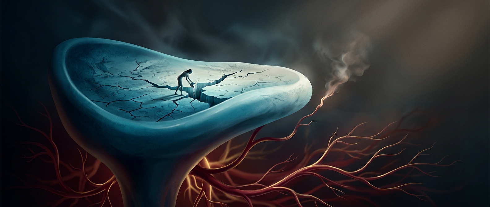
When skin is cut, blood vessels bring oxygen, nutrients, immune cells, and repair signals to the damaged area. Cartilage—the smooth, firm tissue that cushions joints and shapes parts of the ear—has no blood vessels running through it. Its cells therefore receive nutrients mainly by diffusion: molecules slowly pass through the surrounding material rather than arriving in a fresh flow of blood.
That difference matters after an injury. In tissues with a good blood supply, cells involved in repair can reach the wound and begin rebuilding relatively quickly. In cartilage, the few resident cells must work with limited resources, and there is no comparable stream of repair cells entering from nearby vessels. Damage can consequently remain painful or structurally important long after the initial injury.
The location of the injury changes the outlook
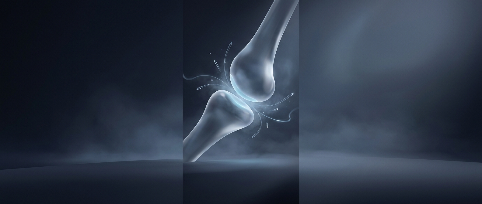
Joint cartilage is especially difficult to repair because it has little capacity to replace damaged tissue with cartilage that matches its original smooth, resilient structure. A defect may instead persist or be filled with repair tissue that does not cushion and glide in quite the same way. The surrounding joint can also keep loading the injured spot with each step or bend, making the consequences noticeable during ordinary movement.
Cartilage is not identical everywhere, and an injury’s depth, size, and location all matter. But the basic constraint is the same: without a direct blood supply, repair depends on slow delivery through surrounding tissue rather than rapid recruitment through vessels. That is why a seemingly small cartilage injury can outlast the moment that caused it—and why returning to activity may require more patience than the pain alone suggests.