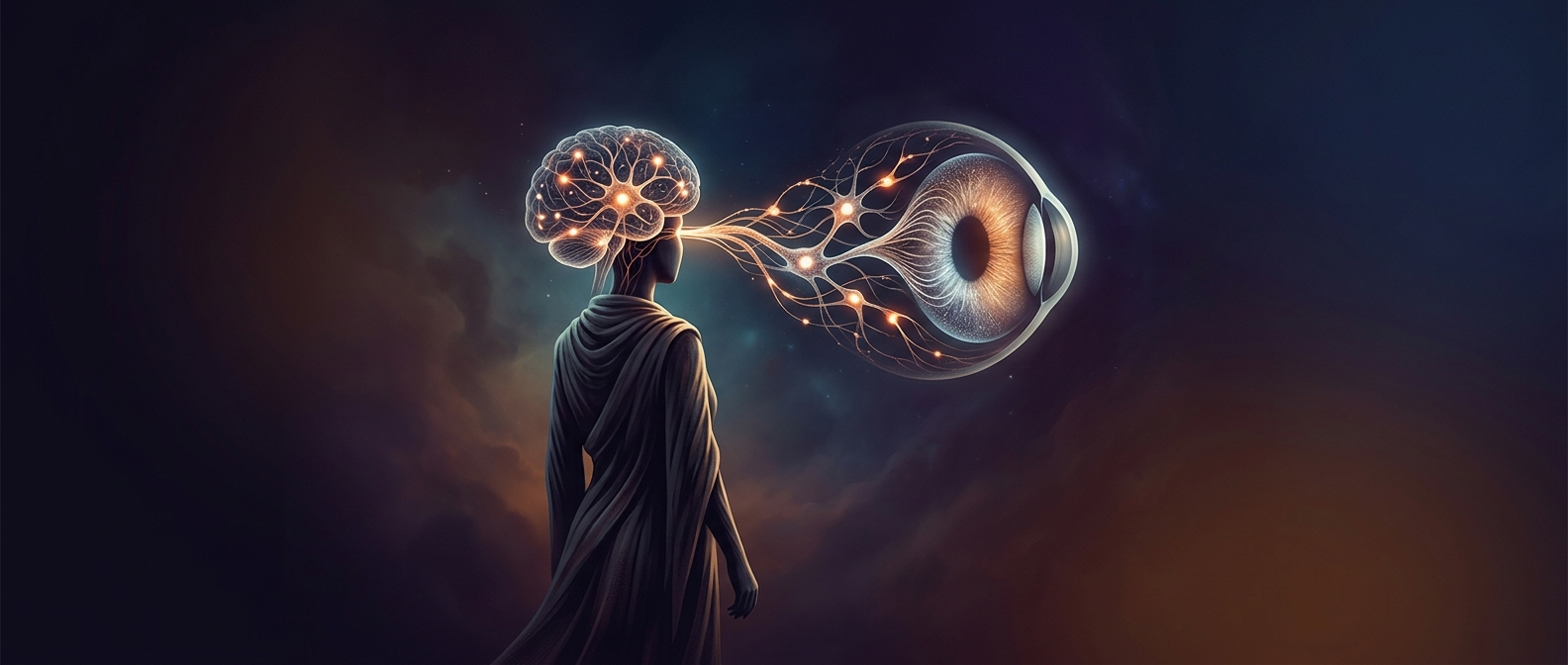
The retina is an outpost of the brain
The retina is the thin, light-sensitive layer lining the back of the eye. During development, it forms from the same embryonic tissue as the brain, and its nerve cells remain connected directly to the brain through the optic nerve. That makes it central nervous system tissue positioned where light enters—not simply a sensor sending raw images inward.
How light becomes a useful signal
Light passes through several retinal layers before reaching photoreceptors: rods, which are especially sensitive in dim conditions, and cones, which support color and fine detail. When light changes the activity of these cells, the retina converts that change into electrical signals. Those signals pass through networks of retinal neurons, including bipolar and horizontal cells, before reaching output cells called ganglion cells. Their long fibers bundle together to form the optic nerve.
The eye begins interpreting the scene
This circuitry does more than relay brightness. Retinal cells compare activity across neighboring regions, helping emphasize edges and changes in contrast. Other pathways respond to features such as color or movement. This early processing helps the brain handle visual information efficiently, rather than receiving a simple point-by-point copy of the light falling on the eye.
The optic nerve carries the retina’s organized signals onward, but the retina is already shaping what those signals contain. In that sense, vision does not begin only when the brain receives the message: part of the first analysis happens at the eye’s inner wall.