Why shattered glass threatened sight
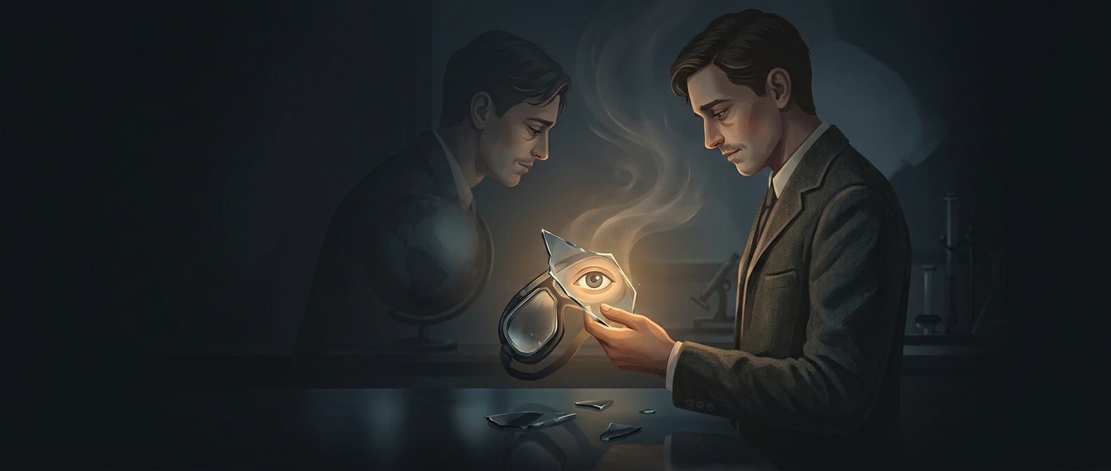
The 1917 Halifax Explosion sent windows bursting across the city. Flying shards cut the corneas—the transparent coverings at the front of the eyes. A cornea is more than a protective surface: its smooth curve helps bend light toward the retina, so a deep cut can leave vision blurred or badly distorted even if the rest of the eye is unharmed.
These injuries were difficult to treat. The tissue is delicate, and a wound that heals unevenly can cloud or warp the cornea. Surgeons had to close damage while preserving the clear, regular surface that sight depends on.
Many similar injuries became a harsh lesson
In the aftermath, doctors encountered large numbers of patients with related eye wounds. That concentration mattered: instead of treating each injury as an isolated puzzle, surgeons could compare what they saw, how they repaired it, and how the eyes healed. Experience with repeated cases helped them refine methods for handling corneal injuries.
The disaster did not make the injuries any less devastating, and it was not a controlled experiment. But the urgent work of treating patients gave eye specialists practical lessons that could inform later surgery. The medical advance came from turning repeated, closely observed injuries into better ways to repair the eye’s most optically sensitive surface.
A grim legacy in clearer vision
The explosion’s glass wounds showed how much the success of eye surgery depends on precision: closing a cut is not enough if the repair leaves the cornea cloudy or misshapen. Halifax’s tragedy therefore became part of the development of corneal surgery—not because disaster was beneficial, but because doctors carried what they learned from its patients into future care.