From a frozen cell to a molecular map
A three-dimensional image of a viral protein is not a photograph in the ordinary sense. In cryo-electron tomography, a beam of electrons passes through a very thin, frozen biological sample. Because electrons scatter as they pass through matter, the detector records patterns that reflect the sample’s internal structure. Each image is a projection: a flattened view of everything along the beam’s path.
The challenge is to turn those overlapping projections into a useful map without losing the proteins’ surroundings. Tilting the sample changes the viewing direction, so the images contain different depth information. Reconstruction software uses the tilt series to estimate where structures sit in three dimensions. The result can reveal viral components in context—among membranes and other cell material—rather than as isolated molecules removed from their setting.
Why the cellular setting matters
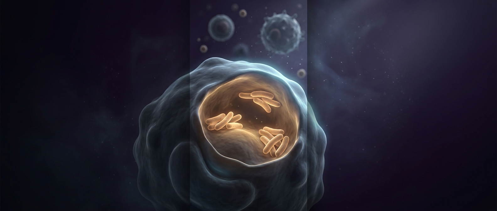
A protein’s shape helps explain what it can do, but its position and neighbors can matter just as much. Seeing a viral protein within a cell can show how it is arranged relative to other structures and offer clues about the steps of infection. That context is difficult to preserve with methods that extract and purify proteins before imaging.
The tradeoff is that a cell is crowded, and the electron beam can damage delicate biological material. Researchers therefore work with very thin regions and limit exposure; this means each image contains some noise and the visible detail is not unlimited. Reconstruction also depends on collecting enough useful angles, which can be difficult when the sample or microscope geometry blocks some views.
The method’s distinctive advantage is context without thawing: scientists can infer the layout of molecular machinery where it exists, while keeping the sample’s frozen structure from being rearranged by liquid water. It turns a series of partial views into a way to ask not only what a viral protein looks like, but where it sits in the cellular scene.