What fossil pigment clues can—and cannot—show
A fossil usually preserves a dinosaur’s shape, not its original paint job. But in some exceptionally preserved specimens, researchers have found melanosomes: microscopic structures that, in living animals, contain melanin, a pigment also found in human skin, hair, and eyes. Their presence can turn a fossil’s dark trace into evidence about how part of an animal may have looked.
Reading color from tiny structures
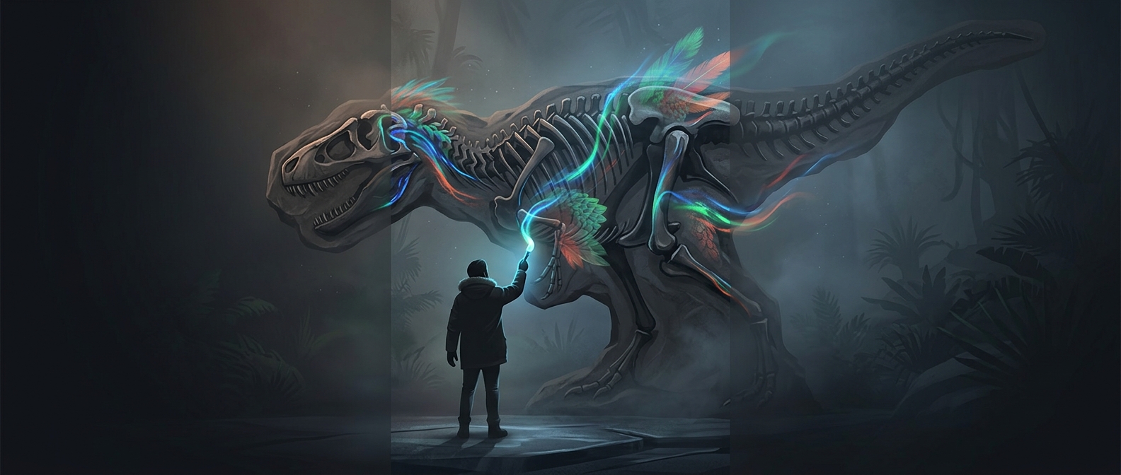
Melanosomes come in different shapes, and their shape and arrangement can help researchers infer whether preserved tissue was dark or reddish-brown. In fossils that retain feathers, scientists can sometimes map these structures across different parts of the body. That offers a patchwork of clues—potentially distinguishing a dark region from a lighter one—rather than a complete portrait.
The inference is not as simple as matching each shape to one exact shade. Fossilization can flatten or alter microscopic structures, and ancient microbes can complicate what researchers see. The evidence is strongest when the structures’ location and pattern fit an animal’s original tissue and survive tests that help separate them from later material.
Why the palette remains incomplete
Melanin is only part of the story. Other pigments and surface features may leave no equally readable microscopic record, and many fossils preserve no useful pigment structures at all. Even a well-preserved patch cannot reveal every color elsewhere on the body, or how the animal’s appearance changed in different light.
So a reconstruction can be evidence-based in one area and speculative in another. A fossil may support a dark feathered back, for example, while leaving the color of the face or underside unknown. That gap is not a failure of the science: it marks exactly where a fossil stops recording and an illustrator has to choose.