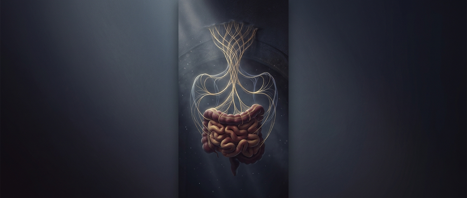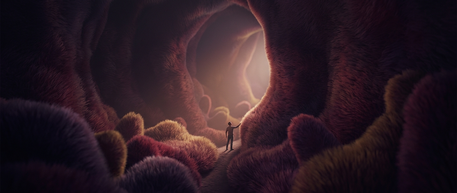
How a flat-looking intestine gains so much working surface
The small intestine does not rely on a smooth tube wall to absorb a meal. Its inner surface is folded into larger ridges and fingerlike villi; in turn, each absorptive cell carries thousands of microscopic projections called microvilli. Together, these layers increase the area available for contact with digested food without requiring a much wider or longer intestine.
Microvilli are not passive bumps. Their outer membranes carry transport proteins that help move particular nutrients into the cells. Some transporters bring in sugars or amino acids alongside sodium; others move nutrients across the cell’s opposite side and onward into circulation. The gradients that power this movement are maintained by cellular pumps, so absorption depends on living tissue, not simply on food touching a surface.
The last steps of digestion happen at the edge
The microvilli form what is often called the brush border. Enzymes attached there finish breaking certain carbohydrates and proteins into small molecules that can be absorbed. This puts digestion and uptake close together: a molecule can be split and then transported across the cell membrane nearby.
Different nutrients take different routes after entering the lining. Many sugars and amino acids pass into tiny blood vessels in the villi. Most dietary fats are packaged into particles that enter lymph vessels first, joining the bloodstream later. The velvet-like texture therefore represents more than extra area: it is a densely equipped boundary where the final chemical cuts and the first steps into the body occur side by side.